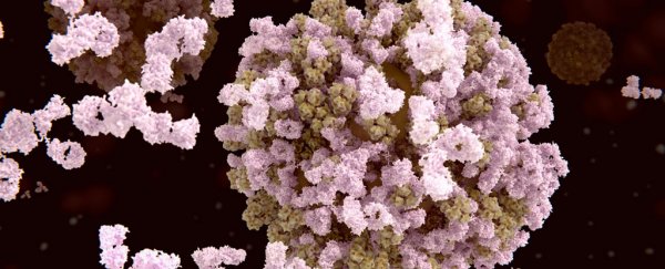
Researchers have discovered that signalling processes between proteins and the immune system are way more complex than previously thought, thanks to a special kind of 'spliced' molecule that's more common than anyone could have predicted.
These molecules were once considered extremely rare, but researchers have found that they actually make up a quarter of the 'flags' that identify toxic or foreign particles in the body – and the discovery could have a huge impact on how we develop vaccines and treat autoimmune conditions.
"It's as if a geographer would tell you they had discovered a new continent, or an astronomer would say they had found a new planet in the Solar System," says systems biologist Michael Stumpf from Imperial College London.
"And just as with those discoveries, we have a lot of exploring to do. This could lead to not only a deeper understanding of how the immune system operates, but also suggests new avenues for therapies and drug and vaccine development."
In a nutshell, our immune system works by identifying things that could be harmful in the body's environment and doing its best to neutralise them.
To do this, it releases proteins called antibodies to take care of antigens, which are the molecules that trigger our immune systems into action.
How does our body recognise antigens? Thanks to a molecular flagging system, known as epitopes. These molecular structures are positioned on the surface of cells, where they identify antigens as antigens.
Conveniently, they're also the right shape and structure for antibodies to bind to them, thereby neutralising any potential threat (hopefully).
Until now, scientists thought that the vast majority of these epitope flags were created in set patterns, with cell machinery effectively 'cutting' fragments out of proteins in a particular sequence, and displaying these in order on the surface of the cell. Wearing house colours, as it were.
But now, thanks to a new cell-mapping technique, scientists have found that as many as a quarter of these flags are actually spliced epitopes – which occur when the protein fragments are displayed on the cell's surface out of order.
In other words, the cells are still wearing house colours, but they've stitched them together chaotically – which hypothetically could make it much harder for the immune system to recognise the patchwork identifier.
Scientists knew these spliced epitopes existed, although they were thought to be uncommon. Not so, according to the new study, which suggests they represent 25 percent of epitopes in terms of abundance, and around 30–40 percent in terms of the diversity of these flagging molecules.
This unexpected volume of spliced identifiers suggests signalling in the immune system carries a lot more 'noise' than scientists were aware of.
And it could be the reason why autoimmune conditions – such as Type 1 diabetes and multiple sclerosis – where the body turns against normal body tissues, are so prevalent.
The discovery is both a blessing and a curse for researchers investigating the immune system. Obviously it's great that we've got a clearer perspective on how many of these spliced epitopes there are, as now we can try to cater to them when developing medical treatments.
But it also means that decoding immune responses just got a lot more difficult.
"The discovery of the importance of spliced peptides could present pros and cons when researching the immune system," says lead researcher Juliane Liepe.
"For example, the discovery could influence new immunotherapies and vaccines by providing new target epitopes for boosting the immune system, but it also means we need to screen for many more epitopes when designing personalised medicine approaches."
The findings are reported in Science.
