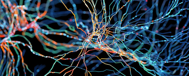
Characterized by shared differences in motor and social behaviors, autism spectrum disorder ( ASD) is also a condition that affects individuals uniquely. Identifying features in the brain that can account for its diverse manifestations and commonalities across all ages has been a goal for scientists seeking to understand its cause.
The latest research, from a team at the University of Rochester in New York, uses advanced scanning techniques to build on previous work on variations in the neurology of people with ASD, offering a closer look at the density and structure of the brain's gray matter.
It's often difficult to do this kind of analysis in living people – so a lot of the existing data we have is based on older post-mortem subjects – but the new image capturing and processing technology means we can now also see how the brain is wired in younger people.
"We've spent many years describing the larger characteristics of brain regions, such as thickness, volume, and curvature," says neuroscientist Zachary Christensen from the University of Rochester.
"However, newer techniques in the field of neuroimaging, for characterizing cells using MRI [ magnetic resonance imaging], unveil new levels of complexity throughout development."
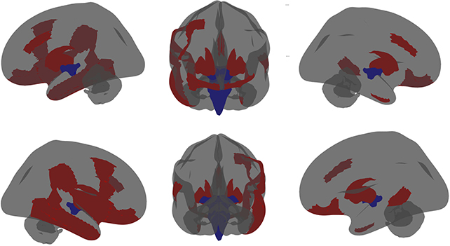
The researchers used a high-contrast form of MRI to build detailed maps of the brains of 142 autistic children, comparing them with images taken from 8,971 controls (children without an autism diagnosis). One set of readings was taken when the volunteers were 9 or 10, with a follow-up set taken a couple of years later.
The comparisons revealed lower neuron densities in certain regions of the cerebral cortex thought to be responsible for our ability to learn, reason, problem solve, and successfully form memories.
In other areas, there was increased neuron density. This was the case in a region called the amygdala, for example, which scientists think helps to process emotions. What's more, when comparing autistic kids to kids with ADHD and anxiety, these differences seemed to be specific to autism.
It's too early to say what these differences in density mean, but they could help explain some of the characteristics of autism. Importantly, the new imaging methods mean we can now track the condition as it develops.
"If characterizing unique deviations in neuron structure in those with autism can be done reliably and with relative ease, that opens a lot of opportunities to characterize how autism develops," says Christensen.
"These measures may be used to identify individuals with autism that could benefit from more specific therapeutic interventions."
It's only relatively recently that we've been able to run non-invasive brain scans with such accuracy and such a high level of detail, and efforts are already underway to follow people with autism over longer periods, to help understand the brain changes that mean they see the world differently.
"It is truly transforming what we know about brain development as we follow this group of children from childhood into early adulthood," says neuroscientist John Foxe, from the University of Rochester.
The research has been published in Autism Research.