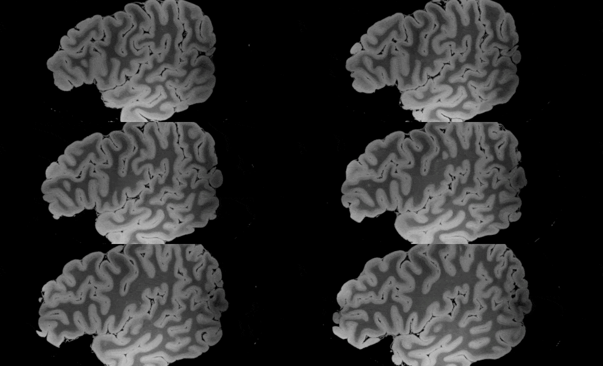
Scientists have produced what looks to be the most detailed magnetic resonance imaging ( MRI) scan ever taken of the human brain anatomy, and are sharing their data with the public.
Thanks to an anonymous deceased patient whose brain was donated to science – and a gargantuan 100 hours of scanning with one of the most advanced MRI machines – the world now has an unprecedented view of the structures that make thought itself possible.
In a new study led by neuroimaging scientist Brian L. Edlow from Massachusetts General Hospital, researchers describe how they recorded their ultra-high resolution MRI dataset of the ex vivo specimen, offering a never-before-seen view of the "three-dimensional neuroanatomy of the human brain".
While their paper has not yet been peer-reviewed, the accomplishment – and the team's decision to share their dataset with anybody who's interested in the research community – is already attracting significant attention, as are high-res videos produced from the scans.
"We haven't seen an entire brain like this," electrical engineer Priti Balchandani from the Icahn School of Medicine at Mount Sinai, who wasn't involved in the study, told Science News.
"It's definitely unprecedented."
The brain came from a 58-year-old woman who was admitted to hospital with fevers, chills, and fatigue, which later developed into breathing problems.
Sadly for the patient, her condition only worsened during her hospital stay, and she died just a fortnight later from respiratory failure caused by viral pneumonia.
While the patient had a history of lymphoma and some other ailments, she had no experience with neurological problems or psychiatric disease, which made her brain a valuable specimen for future neurological research.
After a period of preservation, the organ was transferred to a custom-built, air-tight brain holder made of rugged urethane, specially designed for the experiment's long-duration MRI scan.
The holder was placed in a customised seven Tesla (7T) MRI scanner: a powerful machine offering high levels of magnetic field strength, and only approved by the FDA for use in the US in 2017.
"The overall image quality of MRI improves with higher magnetic field strength," FDA radiologist Robert Ochs explained at the time.
"The added field strength allows for better visualisation of smaller structures and subtle pathologies that may improve disease diagnosis."
In the case of the 7T MRI machine used by Edlow and his team, the researchers were seeking to visualise small structures in the brain specimen at a resolution of 100 micrometres – measuring objects just one tenth of a millimetre in size.
At such minute scales, the researchers explain in their paper, it helps the capture process if the specimen is completely motionless – not to mention, no longer alive.
"Postmortem ex vivo MRI provides significant advantages over in vivo MRI for visualising the microstructural neuroanatomy of the human brain," the authors explain.
"Whereas in vivo MRI acquisitions are constrained by time (i.e. ~hours) and affected by motion, ex vivo MRI can be performed without time constraints (i.e. ~days) and without cardiorespiratory or head motion."
Those benefits, coupled with the capacity for ultra-high resolution imaging across the whole brain, enabled the team to record 8 terabytes of raw data from four separate scan angles, cumulating in approximately 100 hours of total MRI scanning.
In addition to a number of videos of the results, a compressed version of the team's data is now available online for the academic community, with the team saying they "envision a broad range of investigational, educational, and clinical applications for this dataset that have the potential to advance understanding of human brain anatomy in health and disease".
It's early days yet, and it's worth restating that the research still hasn't been published in a peer-reviewed journal; but it's already touted as deeply impactful, and is being integrated into other projects, including a toolbox for deep brain stimulation called Lead DBS.
"Isn't this a beauty?" Lead DBS's Twitter account tweeted about the developments.
"It's hard to predict what exact impact @ComaRecoveryLab's 100 micron 7T postmortem brain will have on the field of brain research, but it is surely going to be tremendous."
The findings are available on the pre-print website bioRxiv.
