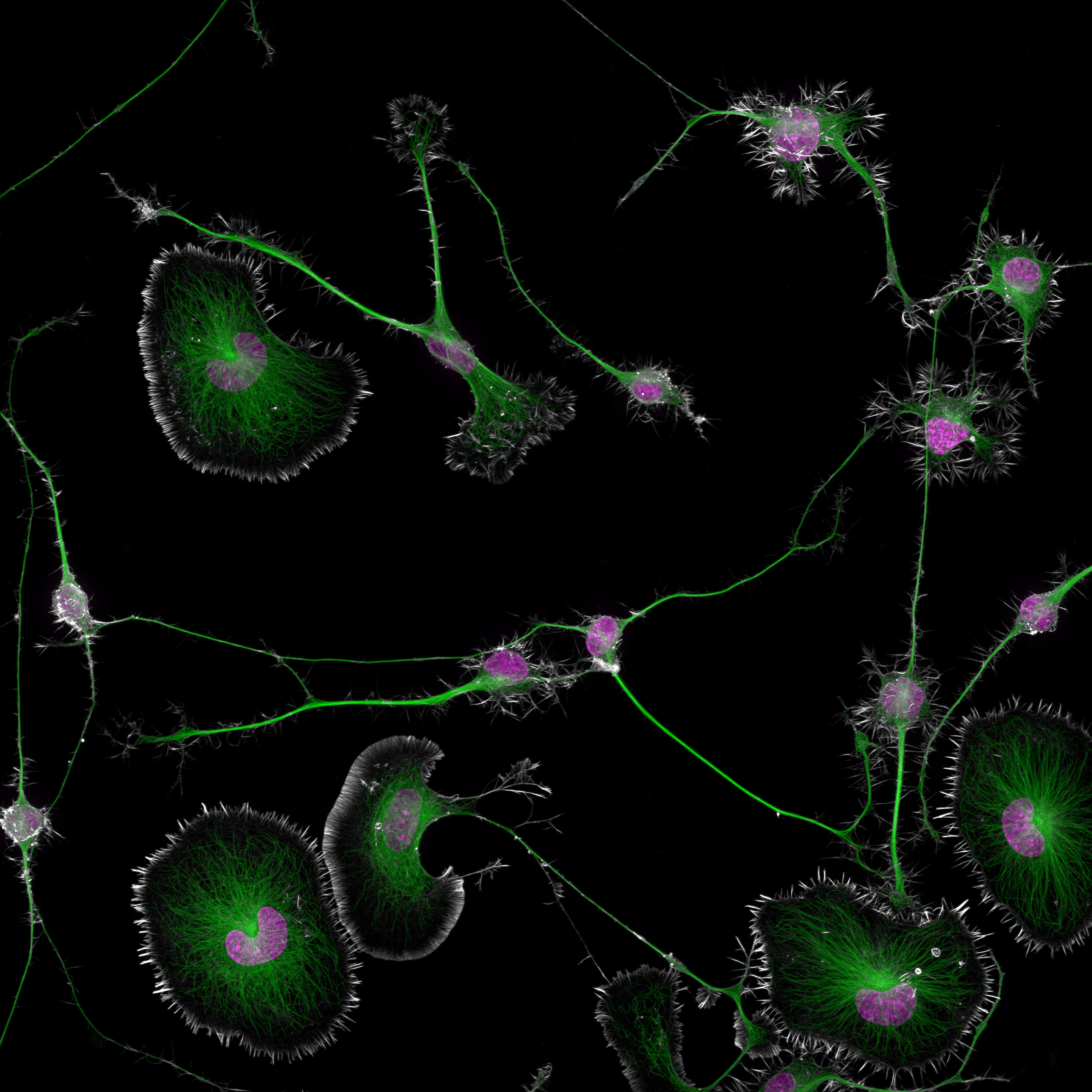
An image more than three months in the making captured the precise moment brain tumor cells from mice interact, winning first place in Nikon's 50th Small World micrography competition.
Augusta University neuroscientist Bruno Cisterna and cell biologist Eric Vitriol stained cellular components to reveal disruptions in support and transport structures that lead to neurodegenerative diseases like Alzheimer's.
"One of the main problems with neurodegenerative diseases is that we don't fully understand what causes them," explains Cisterna. "After three years of research, we finally published our findings."
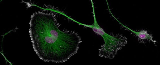
The picture reveals tiny details in two classes of cell; a rounder, less specialized type and longer, more differentiated neuron-like cells. Seen in the image below, the cells' nuclei are stained purple and their cytoskeleton support structures are in green.
Actin and microtubules are the main building blocks of the cytoskeleton, which not only give cells a supporting scaffold but also doubles as a transport system for moving other cellular components around.
Peering closely at these building blocks under the microscope in the two different cell types, Cisterna and colleagues realised that disruptions in a protein linking the two cytoskeleton components together – called profilin 1 (PFN1) – result in damage to the transport system, as seen in neurodegenerative diseases.
Without this molecule, cell components like mitochondria and enzyme storage vessels called lysosomes were whisked around the cell at far greater speeds than normal. And these changes were far more pronounced in the skinny neuron-like cell types where the components were transported across the long branches resembling the axons of nerve cells.
"Enhanced axonal transport has been linked to neurodegenerative diseases like ALS and Alzheimer's disease," Cisterna and team explain in their paper. "Here, we demonstrate that this also may be a consequence of PFN1 loss of function."
Restoring normal cytoskeleton actin and myosin levels allowed the cells to transport their components normally again. This suggests PFN1 regulates the transport system through its interaction with the actin part of the complex.
"To develop effective treatments, we need to figure out the basics first," says Cisterna.
"Our research is crucial for uncovering this knowledge and ultimately finding a cure. Differentiated cells could be used to study how mutations or toxic proteins that cause Alzheimer's or ALS alter neuronal morphology, as well as to screen potential drugs or gene therapies aimed at protecting neurons or restoring their function."
These results highlight how scientific imaging can help expose biological mysteries.
"Sometimes, we overlook the tiny details of the world around us," says Nikon Instruments senior manager Eric Flem. "Nikon Small World serves as a reminder to pause, appreciate the power and beauty of the little things, and to cultivate a deeper curiosity to explore and question."
This research was published in the Journal of Cell Biology and you can see a whole realm of other microscopic wonders here.