A few years ago, researchers were finding that psychology research was being affected by something WEIRD – an oversampling from Western, Educated, Industrialised, Rich, and Democratic communities.
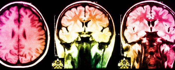
Now it seems neurology suffers from a similar issue, with a recent study revealing that low diversity among volunteers in neuroimaging research projects risks warping our impression of what a normal growing brain looks like.
Researchers from the US used a database on paediatric neurological development to determine whether the particular mix of cultural demographics made any real difference to representative measurements of brain structures.
There's no shortage of studies on brain development in children and adolescents, providing scientists with a wealth of statistics on the average thickness, density, mass, or surface area of some zone or node or lobe of the brain at a given age.
These numbers contribute to a wider understanding of what a normal nervous system should look like as it develops.
While some researchers go to great lengths to match the mix of their sample with the population they're describing, not all have such fortune or tenacity.
Sometimes scientists are forced to take what they can get as far as volunteers go. Time and funding can limit the diversity of subjects willing to take time out of their schedule to have their brains – or those of their children – zipped through a scanner.
Most subjects also come from virtually right outside the research facility's front door, meaning a good many brain scans are done on college and university students or their families.
That means there's an overabundance of certain demographics among the pool of data.
The researchers found children from a European Caucasian background were underrepresented in the database, with more having highly educated parents, and slightly higher family incomes.
To see if this biased dataset made an appreciable difference to how researchers might catalogue brain development, the team constructed a sample made up of 1162 individuals that represented the balance of racial, socioeconomic, and educational features across the US.
They then compared average developmental milestones within this weighted group with figures from the biased population, and found a significant difference in what could be considered 'normal'.
When the sample was manipulated to look more like the general US population, the surface areas of specific lobes grew at a slightly faster rate at an earlier age before tapering off in adolescence.
The timing of development also varied between the samples; without weighting, the sample suggested the parietal lobe reached peak surface area first in most children, followed by the temporal, occipital, and frontal lobes.
When skewed to reflect the wider community, the results showed the occipital and parietal lobes peaking, followed by the temporal lobes, and then the frontal.
Similar differences were found in other patterns of growth, demonstrating that the statistical landmarks defining what was physiologically average shifted significantly with the sample's diversity.
The results say nothing on how these changes manifest in terms of psychological or social development.
But they are a reminder of how we should be careful not avoid reading too much into brain scan results based on samples with limited cultural diversity.
Similar words of caution have been argued before in psychology, where a predominance of papers based on white, affluent Americans risked painting a biased picture of what constitutes a mentally healthy individual.
It might not demand a bonfire for developmental neurology text books, but it should be a wake up call when it comes to the finer details of data based on small, non-representative samples.
Incorporating population science into neuroimaging research would go a long way to addressing the problem, although the same challenges will no doubt continue to plague researchers seeking a diverse population to recruit from.
Meanwhile, a good dose of critical thinking never goes astray when it comes to studies on subjects as complex as culture and the human brain.
This research was published in Nature Communications.
