Death, to put it mildly, is a rather inconvenient event for a living brain. The cascade of effects that arises as oxygen vanishes sweeps like a tide down to the very way our cells transcribe and translate our DNA, scrambling in a last-ditch attempt to keep the lights on.
A comparison of post-mortem brain tissue and samples taken from living patients has revealed for the first time significant differences in the way strands of RNA are modified, exposing new potential targets for disease diagnosis and treatment.
Researchers from the Icahn School of Medicine at Mount Sinai in New York focused on the way specific base codes of adenosine (A) are swapped for a completely different base, inosine (I), in messenger RNA.
"Until now, the investigation of A-to-I editing and its biological significance in the mammalian brain has been restricted to the analysis of postmortem tissues," says genomicist Michael Breen.
"By using fresh samples from living individuals, we were able to uncover significant differences in RNA editing activity that previous studies, relying only on postmortem samples, may have overlooked."
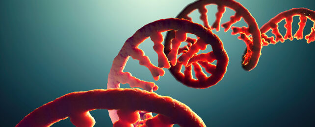
To turn the genes encoded by double-stranded helixes of DNA into functional proteins, biology has to copy their sequences into a subtly different format based instead on RNA. These 'messengers' can then be translated into proteins by other RNA structures that piggyback the amino acid building blocks.
Billions of years of evolution has taken advantage of this intermediary transcription and translation service to virtually add in a whole new library of proteins. Like a rogue editor rewording manuscripts to serve entirely new purposes, cells can tweak a gene's messenger RNA to meet entirely different needs.
Some species – most notably types of cephalopod – take RNA editing to a whole new level, rewriting their brain's own genetic instructions as the occasion calls for it.
In vertebrates such as ourselves, the removal of an amino group, or 'deamination' of adenosine turns it into inosine – a base similar to the base guanine (G) – typically resulting in a very different end product to the one encoded for in the DNA's library of genes.
This A-to-I base swap is accomplished by the adenosine deaminase acting on RNA (ADAR) family of enzymes, which play critical roles in shaping a range of different tissues, including those in the brain.
The process is so critical, in fact, that errors in the editing process can result in a variety of neurological disorders. To determine precisely how edits to specific transcribed genes develop into life-threatening conditions, researchers have analyzed specimens collected post-mortem.
As convenient as these samples might be to collect, they suffer a major drawback.
"We hypothesized that molecular responses to postmortem-induced hypoxic and immune responses can significantly alter the landscape of A-to-I editing," says the study's lead author Miguel Rodríguez de los Santos, a molecular biologist at Mount Sinai.
"This can lead to misunderstandings about RNA editing in the brain if we only study postmortem tissues."
Sure enough, samples of brain tissue obtained from living patients during the surgical placement of deep brain stimulation electrodes revealed major differences in the activity of two kinds of ADAR enzymes, as well as the sites they acted upon.
The team's analysis distinguished in excess of 72,000 locations on RNA strands where A-to-I editing occurred more often in specimens from the recently deceased, compared with those collected from a living patient.
There were hundreds of sites where the opposite occurred, however, where the editing process was more prolific in the samples from living brains. While some of the sites had known functions in brain plasticity, many require further investigation to understand the mechanisms at play.
"It is critical to note that our findings do not negate but instead provide missing context for using postmortem brain tissues in researching A-to-I regulation," says co-senior author Alexander Charney, a physician-scientist at Mount Sinai.
"Understanding these differences helps improve our knowledge of brain function and disease through the lens of RNA editing modifications, which can potentially lead to better diagnostic and therapeutic approaches."
This research was published in Nature Communications.