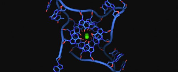
Two thin strands wound together in a spiraling helix: This is the iconic shape of a DNA molecule. But sometimes, DNA can form a rare quadruple-helix, and this odd structure may play a role in diseases like cancer.
Not much is known about these four-stranded DNA, known as G-quadruplexes — but now, scientists have developed a new way to detect these odd molecules and observe how they behave in living cells.
In a new study, published January 8 in the journal Nature Communications, the team described how certain proteins cause the G-quadruplex to unravel; in the future, their work could lead to new drugs that grab hold of quadruple-helix DNA and disrupt its activity.
Drugs could intervene, for instance, when the odd DNA contributes to cancerous tumor growth.
"Evidence has been mounting that G-quadruplexes play an important role in a wide variety of processes vital for life, and in a range of diseases," study author Ben Lewis, from the Department of Chemistry at Imperial College London, said in a statement.
Related: 7 diseases you can learn about from a genetic test
In general, G-quadruplexes crop up in cancer cells at much higher rates than healthy cells, according to the statement.
Various studies have linked the presence of four-stranded DNA to the rapid division of cancer cells, a process that leads to tumor growth; so scientists hypothesized that targeting the weird DNA with drugs could slow or stop this unbridled cell division. Some studies already support this idea.
"But the missing link has been imaging this structure directly in living cells," Lewis said. In other words, scientists needed a better way to watch these DNA molecules in action.
The new study begins to fill in that missing knowledge.
G-quadruplexes can form either when one double-stranded DNA molecule folds over on itself or when multiple DNA strands link up at a single nucleic acid, known as guanine — one of the building blocks of DNA, according to Discover Magazine.
To spot this funky DNA in cells, the team used a chemical called DAOTA-M2, which emits a fluorescent light when it binds to G-quadruplexes. Rather than only measuring the brightness of the light, which varies depending on the concentration of DNA molecules, the team also tracked how long the light shone.
Tracking how long the light lingered helped the team see how different molecules interacted with the four-stranded DNA in living cells.
When a molecule latched onto the DNA strand, it would displace the glowing DAOTA-M2, causing the light to go out faster than if the chemical had remained in place. Using these methods, the team identified two proteins, called helicases, that unwind the strands of four-stranded DNA and jumpstart the process of breaking them down.
They also identified other molecules that bind to the DNA; future studies on these molecular interactions could help scientists design drugs that bind to the DNA.
"Many researchers have been interested in the potential of G-quadruplex-binding molecules as potential drugs for diseases such as cancers," Ramon Vilar, a professor of medicinal inorganic chemistry at Imperial, said in the statement.
"Our method will help to progress our understanding of these potential new drugs."
Related content:
Genetics by the numbers: 10 tantalizing tales
7 odd things that raise your risk of cancer (and 1 that doesn't)
Science experiment for kids: Seeing your DNA
This article was originally published by Live Science. Read the original article here.
