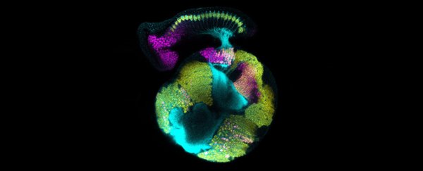
Pop quiz: what kind of cells form a human brain? If you said neurons, you get part marks – the brain wouldn't be half the organ it is if not for helper cells called glia.
But don't feel too bad. In the past, researchers have also focussed primarily on the neuron in the starring role of brain development in a growing embryo. Now it turns out those glial cells should be getting a bigger cut of the glory.
A small team of researchers led by scientists from New York University turned to an oft-used model of animal development to study how a nervous system grows – the humble fruit fly.
Specifically, the researchers studied the visual system of flies called Drosophila in order to see how dividing cells became circuits tasked with mapping the light that falls on sensory nerves.
Usually this kind of process would be associated solely with cells that are already known to respond to chemical changes by passing on a signal – aka nerve cells.
Instead, the researchers found cells that usually stay backstage have a rather active role to play in the process.
"The results lead us to revise the often neuro-centric view of brain development to now appreciate the contributions for non-neuronal cells such as glia," says researcher Vilaiwan Fernandes from New York University.
Glia come in a variety of forms, but all of them are dedicated to helping neurons do their all-important job of transmitting waves of electrochemistry.
This support crew isn't a small team, either, making up at least half of the total number of cells in the brain.
The name itself comes from the Greek word for glue, but in recent years it's become clear that they do far more than form a scaffold to hold nerve cells together, such as mopping up stray and unwanted chemicals, providing access to nutrients, and offering protection.
In spite of being recognised for its multitude of side-jobs, researchers have still viewed a distinction between the active circuitry of neurons and the simple servitude of the glia.
This new research suggests the boundary is a lot fuzzier than most had assumed.
"Indeed, our study found that fundamental questions in brain development with regard to the timing, identity, and coordination of nerve cell birth can only be understood when the glial contribution is accounted for," says Fernandes.
Brain development in even relatively simple organisms like fruit flies requires strict coordination in adjusting numbers and connections of cells accordingly.
While there's a world of difference between humans and flies, neurologically both are made up of mini-circuits that build together to perform a task, such as collecting light and turning it into a useful map.
Rather than this coordination coming from the neurons, the researchers found it's the glia that oversee the construction, boosting noisy signals and sending insulin-like messages between the retina and the emerging optical sections of the brain to induce specific changes.
It wasn't entirely clear why the glial cells have taken up such an important role, especially given nerve cells already connect the different structures and could pass on the signals themselves.
The researchers hypothesised that the glia helped ensure the necessary changes in the optical lobes of the brain only took place after the right pathways had been established.
They tested this by passing on the glial cell's insulin-like signals in developing flies prematurely, demonstrating the glia's role in getting the timing of specific changes right.
"By acting as a signaling intermediary, glia exert precise control over not only when and where a neuron is born, but also the type of neuron it will develop into," says senior researcher Claude Desplan.
This is just a single example of glia directing neurological development of one part of the nervous system.
But it sets up the possibility that what we've considered to just be nanny cells could be more like the principals in certain areas of brain development.
It's time our brain's non-acting cast stepped out and took a bow.
This research was published in Science.
