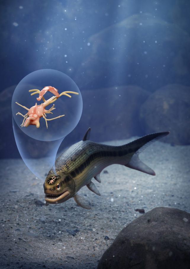
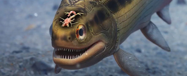
Paleontologist Matt Friedman was surprised to discover a remarkably detailed 319-million-year-old fish brain fossil while testing out micro-CT scans for a broader project.
"It had all these features, and I said to myself, 'Is this really a brain that I'm looking at?'" says Friedman from University of Michigan.
"So, I zoomed in on that region of the skull to make a second, higher-resolution scan, and it was very clear that that's exactly what it had to be. And it was only because this was such an unambiguous example that we decided to take it further."
Usually, the only remaining traces of such ancient life are from more easily preserved hard parts of animals, like their bones, since soft tissues degrade quickly.
But in this case, a dense mineral, possibly pyrite, seeped in and replaced tissue that had likely been preserved for longer in a low-oxygen environment. This allowed scans to pick up what look like cranial nerve and soft tissue details of the small fish, Coccocephalus wildi.
The ancient specimen is the only one of its kind, so despite having been in the hands of researchers since it was first described in 1925, this feature remained hidden as scientists would not risk invasive methods of investigation.
"Here we've found remarkable preservation in a fossil examined several times before by multiple people over the past century," explains Friedman.
"But because we have these new tools for looking inside of fossils, it reveals another layer of information to us."
This prehistoric estuary fish likely hunted insects, small crustaceans, and cephalopods, chasing them with fins supported by bony rods called rays.
Ray-finned fish, subclass Actinopterygii, make up over half of all living backboned animals alive today, including tuna and seahorses, and 96 percent of all fish.
This group split from lobe-finned fishes – some of which eventually became our own ancestors – about 450 million years ago. C. wildi then took its own evolutionary path from the groups of fishes still living today around tens of millions of years ago.
"Analyses place this taxon outside the group containing all living ray-finned fish species," University of Michigan paleontologist Rodrigo Figueroa and colleagues write in their paper.
"Details of the brain structure in Coccocephalus therefore have implications for interpretations of neural morphology during the early evolutionary stages of a major vertebrate lineage."

Some brain features would have been lost to decay and the preservation process, but the team could still make out specific morphological details. This allowed them to see that the way this prehistoric forebrain developed was more like ours than the rest of the living ray-finned fishes alive today.
"Unlike all living ray-finned fishes, the brain of Coccocephalus folds inward," notes Friedman. "So, this fossil is capturing a time before that signature feature of ray-finned fish brains evolved. This provides us with some constraints on when this trait evolved – something that we did not have a good handle on before the new data on Coccocephalus."
This inward fold is known as an evaginated forebrain – like in us, the two brain hemispheres end up embracing a hollow space like a 'c' and its mirror image joined together. By comparison, everted forebrains seen in still-living ray-finned fishes have two puffed-up lobes instead, with only a thin crevice between them.
The researchers are keen to scan other fish fossils in the museum's collections to see what other signs of soft tissue may be hiding within.
"An important conclusion is that these kinds of soft parts can be preserved, and they may be preserved in fossils that we've had for a long time – this is a fossil that's been known for over 100 years," says Friedman.
"That's why holding onto the physical specimens is so important. Because who knows, in 100 years, what people might be able to do with the fossils in our collections now."
This research was published in Nature.
