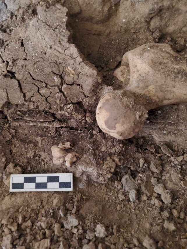
If it weren't for the careful eyes of an excavator, working at an ancient underground tomb in Egypt, archaeologists may never have found it: a lonely tooth, nestled in the curve of a worn-down pelvis.
At first, site supervisor and archaeologist Melinda King Wetzel thought she was looking at a fetus from the time of the Egyptian pharaohs.
But when she showed the grave to the bioarchaeological director of the site, Gretchen Dabbs, the discovery turned out to be even rarer in nature.
Along with another site supervisor, Anna Stevens, Wetzel and Dabbs claim to have found the oldest evidence of a mature ovarian teratoma, or germ cell tumor.
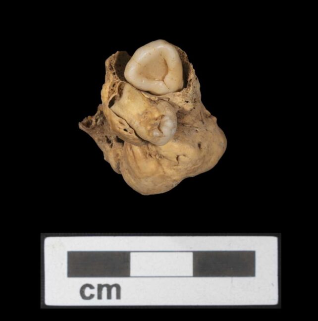
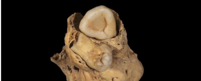
Today, the mass looks like a calcified clump of disorganized and fully formed tissues, like bone and teeth.
It measures roughly 3 by 2 centimeters (0.8 by 1.2 inches) in dimension and dates back to the mid-14th century BCE.
Researchers say it adds "considerable temporal and geographical depth to our understanding of this condition in the past."
Wetzel, Dabbs, and Stevens, who all hail from different companies and universities, have been working together at this archaeological site, on the eastern banks of the Nile River, for years now, as part of the Amarna Project.
This is a long-term, ongoing dig that seeks to uncover the cemeteries of normal folk buried near what was once the capital city of the Pharaoh Akhenaten, established from 1345 BCE.
The young female skeleton with the ovarian tumor was found buried in a multi-chambered tomb at Amarna's North Desert Cemetery and was probably 18 to 21 years of age when she died.
She was buried with her hands positioned over her pelvis and wrapped in a way that was common for other non-elite Amarna cemeteries. That said, she had more jewelry on her than other bodies nearby.
Within the ring of her pelvic bone, however, lay a different sort of jewel.

Teratomas are rare types of germ cell tumors that are usually benign, although they can come with symptoms such as abdominal pain or infertility.
They are very rarely found in archaeology. In fact, this is only the fifth case of its kind to be uncovered by archaeologists and the only one from Egypt.
The tumor is several centuries older than the other ancient teratomas previously discovered by archaeologists in Spain, France, Peru and Portugal.
Initially, when Wetzel saw the tooth, she thought it was alone. But upon closer inspection, she and her colleagues noticed another empty slot on the calcified mass.
Further excavations around the female skeleton revealed a second tooth near the top of the leg bone, still within the pelvic cavity. It fit into the empty slot nicely, which led archaeologists to conclude that this second tooth used to be part of the tumor but had separated during decomposition.

Both teeth are covered in full, "if somewhat disfigured", enamel crowns, according to the authors. They also show partially formed root structures at their cemento-enamel base.
This is much too developed for an early stage fetus and it strongly suggests the mass is not an unborn offspring but a tumor.
"Without the careful excavation and recording of the teratoma in situ," writes the team of archaeologists, "it is more than likely the isolated tooth would have been, at least initially, identified as an intrusive element from a different individual or possibly as evidence of an additional burial within the tomb if the tooth was not able to be associated with the other individuals buried in the same tomb."
This particular individual, the researchers say, had a gold ring on her left hand that sat close to the tumor. It was illustrated with an image of Bes, an ancient Egyptian deity associated with fertility and protection.
While just a hypothesis, the authors say it is conceivable that this ring position was purposeful and that the Bes ring might have been used to address the pain felt in this part of the body or "perceived issues of infertility".
The study was published in the International Journal of Paleopathology.
