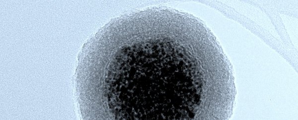
For the first time, researchers have found that nanoparticles shaped like rods and worms are far more effective at travelling through cells and specific barriers like the nucleus than spherical ones.
This is big news for the development of new drug delivery systems, because one of the main struggles for researchers in the field is getting drug-toting nanoparticles into the correct part of the cell in the first place.
"We were able to show for the first time that nanoparticles shaped like rods and worms were more effective than spherical nanoparticles at traversing intracellular barriers and this enabled them to get all the way into the nucleus of the cell," says lead author Elizabeth Hinde, from the University of New South Wales (UNSW) in Australia.
The team applied a new fluorescent microscopy method to standard drug delivery, which allowed them to track the movement of nanoparticles of different shapes through a single cancer cell.
They were then able to pinpoint where the drug was being released, and how it spread throughout the cell.
"You need to know how things arrive at their final destination in order to target them there," says Hinde. "Now we have a tool to track this incredible journey to the centre of the cell. It means other research groups can use this to assess their nanoparticles and drug delivery systems."
"They'll be able to work out how to tailor their particles to reach the nucleus or other structures in the cell, and gauge where the cargo is being dropped off. This wasn't possible before," she adds.
But let's step back a bit and talk about nanoparticles. Nanoparticles are defined as particles between 1 and 100 nanometres in diameter, and they're increasingly being used to transport drugs to specific areas of the body, kill cancer cells, and allow researchers to image the body.
The problem is, nanoparticles are not perfect. Although many nanoparticle technologies seem promising, some fall flat on actually making it into the correct area of the cell, because cells are actually really cramped and it can be hard to get there.
Hinde's team looked at four types of nanoparticle shapes: micelles and vesicles, which are two types of spheres; as well as rod- and worm-shaped nanoparticles.
When the researchers used doxorubicin (a cancer drug) in the different shaped nanoparticles, the rod and worms passively entered the nucleus without any issues. The spherical ones, on the other hand, were stuck outside the nucleus.
Getting through the nuclear membrane and into the nucleus is important for increasing the toxicity of cancer cells, so rods and worms came out on top.
"Cancer cells have different internal architecture than healthy cells," says Hinde.
"If we can fine-tune the dimensions of these rod-shaped nanoparticles, so they only pass through the cellular barriers in cancer cells and not healthy ones, we can reduce some of the side effects of chemotherapies."
The researchers aren't just hoping to use this on cancer drugs though - this new technology has the ability to change the way we analyse nanoparticles generally.
"The impact for the field is huge," says Justin Gooding, one of the researchers from UNSW.
"It gives us the ability to look inside the cell, see what the particles are doing, and design them to do exactly what we want them to do."
The best thing for the researchers is that this technique can be performed using current equipment in most labs - so no new machines are required.
"And this isn't just thanks to the microscope, but the information and data we can extract from the new analysis procedures we've developed," says Gooding.
"People are going to see, suddenly, that they can get all sorts of new information about their particles."
We're excited to see where this will lead.
The research has been published in Nature Nanotechnology.
UNSW Science is a sponsor of ScienceAlert. Find out more about their world-leading research.
