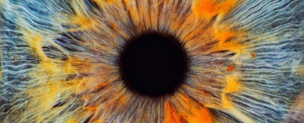
Scientists have momentarily restored a faint twinkle of life to dying cells in the human eye.
In order to better understand the way nerve cells succumb to a lack of oxygen, a team of US researchers measured activity in mouse and human retinal cells soon after their death.
Amazingly, with a few tweaks to the tissue's environment, they were able to revive the cells' ability to communicate hours later.
When stimulated by light, the postmortem retinas were shown to emit specific electrical signals, known as b-waves.
These waves are also seen in living retinas, and they indicate communication between all the layers of macular cells that allow us to see.
It's the first time deceased human donor eyes have ever responded to light in this way, and it has some experts questioning the irreversible nature of death in the central nervous system.
"We were able to wake up photoreceptor cells in the human macula, which is the part of the retina responsible for our central vision and our ability to see fine detail and color," explains biomedical scientist Fatima Abbas from the University of Utah.
"In eyes obtained up to five hours after an organ donor's death, these cells responded to bright light, colored lights, and even very dim flashes of light."
After death, it's possible to save some organs in the human body for transplantation. But after circulation ceases, the central nervous system as a whole stops responding far too quickly for any form of long-term recovery.
Yet not all types of neuron fail at the same rate. Different regions and different types of cell have different survival mechanisms, making the whole brain-death issue a lot more complicated.
Learning how select tissues in the nervous system cope with a loss of oxygen could teach us a thing or two about recovering lost brain functions.
Researchers have had some luck already. In 2018, scientists at Yale University made headlines when they kept pig brains alive for as long as 36 hours after death.
Four hours postmortem, they were even able to revive a small response, though nothing organized or global that could be measured by an electroencephalogram (EEG).
The feats were achieved by stalling the rapid degradation of mammalian neurons, using artificial blood, heaters, and pumps to restore circulation of oxygen and nutrients.
A similar technique now appears possible in mice and human eyes, which is the only extruding part of the nervous system.
By restoring oxygenation and some nutrients to organ donor eyes, researchers at the University of Utah and Scripps Research were able to trigger synchronous activity among neurons after death.
"We were able to make the retinal cells talk to each other, the way they do in the living eye to mediate human vision," says visual scientist Frans Vinberg from the University of Utah.
"Past studies have restored very limited electrical activity in organ donor eyes, but this has never been achieved in the macula, and never to the extent we have now demonstrated."
Initially, the experiments showed retinal cells continued to react to light for up to five hours after death. Yet the crucial inter-cellular b-wave signals quickly dropped off, apparently due to the loss of oxygen.
Even when retinal tissue is carefully protected from oxygen deprivation, the researchers were unable to completely restore robust b-waves.
The temporary revival of the retinal cells doesn't mean the donor eyeballs could 'see', of course. Higher visual centers in the brain are needed to revive full visual sensation and perception.
Nonetheless, some definitions of 'brain death' require a loss of synchronous activity among neurons. If that definition is accepted, then the human retinas in the current study were not yet fully dead.
"Since the retina is part of the CNS, our restoration of the b-wave in this study raises the question of whether brain death, as it is currently defined, is truly irreversible," the authors write.
If specialized neurons, known as photoreceptors, can be revived to a certain extent, then it offers hope for future transplantations that could help restore vision in those with eye disease.
That day, however, is still a long ways off. Transplanted cells and patches of a donor retina would need to somehow be seamlessly integrated into existing retinal circuits, which is a daunting challenge scientists are already trying to tackle.
In the meantime, donor eyes and animal models will have to do, and testing for b-waves could be a good way to determine if a retinal graft is viable or not.
"The scientific community can now study human vision in ways that just aren't possible with laboratory animals," says Vinberg.
"We hope this will motivate organ donor societies, organ donors, and eye banks by helping them understand the exciting new possibilities this type of research offers."
The study was published in Nature.
