The human brain is a ridiculously complex organ that doesn't give up its secrets easily. Thanks to advances in imaging technology, hidden forms and functions of neurological anatomy continue to emerge, from novel kinds of nerve cells to entirely new nubs of tissue.
Now researchers from the University of Copenhagen and University
of Rochester have identified a layer of tissue that helps protect our gray and white matter, one that hasn't been distinguished before.
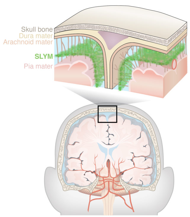
Only a few cells thick, this membrane seems to play a role in mediating the exchange of small, dissolved substances between compartments in the brain. It also appears to be the home base of brain-specific immune cells, not to mention assisting in the brain's waste-removal (glymphatic) system.
University of Copenhagen molecular biologist Kjeld Møllgård and colleagues have called their discovery the Subarachnoid LYmphatic-like Membrane (SLYM). While much of their research on this structure is so far from mice, using two-photon microscopy and dissections, they have confirmed the SLYM's presence in an adult human brain too.
The SLYM lies between two other membranes protecting the brain. It divides our brain fluid space in two, bringing the total number of known membranes encasing our brain up to four. It appears to act as a barrier for molecules in our brain fluid that are larger than around 3 kilodaltons; comparable to an extremely small protein.

Unlike the rest of our body, our central nervous system does not have lymphatic (immune) vessels and is considered immune privileged – a term that refers to sites in our bodies where immune responses are highly controlled, such as our eyes and testes.
So the team suspects cerebrospinal fluid may pick up some of the immune system's role in the brain. The presence of the SLYM could explain how this works.
"The discovery of a new anatomic structure that segregates and helps control the flow of cerebrospinal fluid (CSF) in and around the brain now provides us much greater appreciation of the sophisticated role that CSF plays not only in transporting and removing waste from the brain, but also in supporting its immune defenses," says University of Rochester neuroscientist Maiken Nedergaard.
Møllgård and team found several types of immune cells – including myeloid cells and macrophages – camping out in the SLYM, keeping surveillance over the brain. In mice, the types of cells changed in response to swelling and natural aging, suggesting this site may play an important part in disease pathologies.
The SLYM shares molecular markers with the mesothelial membrane that lines the rest of our organs, encasing their blood vessels and storing immune cells. So the researchers propose that the SLYM is the brain's mesothelium, lining the blood vessels in the cavity between the brain and skull.
Mesothelium also plays a role of lubricant between organs that slide against each other.
"Physiological pulsations induced by the cardiovascular system, respiration, and positional changes of the head are constantly shifting the brain within the cranial cavity," the researchers explain in their paper. "SLYM may, like other mesothelial membranes, reduce friction between the brain and skull during such movements."
Tears in the SLYM may explain some of the long term symptoms of traumatic brain injury, Møllgård and team speculate. Disruption of this barrier would allow immune cells from the skull direct access into the brain, cells that are not calibrated for brain conditions. This could explain ongoing inflammation.
The flow of waste out of the brain can also continue to be suppressed for a long period after brain injury, and altered flow patterns of the cerebrospinal fluid because of the membrane's rupture may explain this.
As this extra layer of brain armor has only just been discovered, there's still a lot to work out. The researchers question if this tissue may also be involved in a more general immunity of the central nervous system and therefore play a role in associated diseases like multiple sclerosis.
"We conclude that SLYM fulfills the characteristics of a mesothelium by acting as an immune barrier that prevents exchange of small solutes between the outer and inner subarachnoid space compartments and by covering blood vessels in the subarachnoid space," write Møllgård and colleagues.
This research was published in Science.
