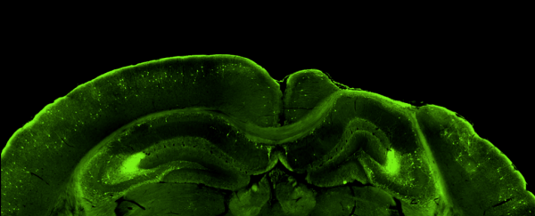
We know the brain changes after traumatic injury, and now we have maps from mice showing what that change looks like.
A team of scientists has traced connections between nerve cells throughout the entire brain of mice, showing that distant parts of the brain become disconnected after a head injury.
The stunning visualizations of brain-wide connectivity could help scientists understand how a traumatic brain injury, or TBI, alters cross-talk between different cells and brain regions, first in mice and then in humans.
"We've known for a long time that the communication between different brain cells can change very dramatically after an injury," says neuroscientist and study author Robert Hunt of the University of California, Irvine (UCI), who envisaged the project a decade ago.
"But we haven't been able to see what happens in the whole brain until now."
There's still so much we don't fully understand about traumatic brain injuries, which can leave people with lifelong disabilities, feeling like shadows of their former selves and almost unrecognizable to family.
A TBI results when a blow to the head – often from a fall, car accident, sports collision, or physical assault – sends the brain ricocheting around inside the skull, causing lasting damage.
Repeated head traumas leading to a severe condition known as chronic traumatic encephalopathy have been well-documented in professional athletes. But even 'mild' head knocks called concussions can manifest damage years later, recent research shows.
No two head injuries are usually the same, making them challenging to study, although there are common symptoms: memory problems, communication difficulties, attention deficits, depression, and emotional instability, to name but a few.
However, linking behavioral, emotional, and brain function changes to changes in specific brain cells or wider neural networks is one of the important tasks at hand, as researchers hope to better understand how brain damage develops and if its onset could be prevented.
In this study, Hunt and the team, led by fellow neuroscientist and UCI researcher Jan Frankowski, devised a few new and improved techniques to map connections between nerve cells across the entire brain in a mouse model replicating TBI using a dazzling array of laser-illuminated fluorescent tags.
By that time, iDISCO was well established and we modified the clearing protocols to achieve strong immunoslabeling throughout an entire injured brain, without separating the brains into two hemispheres as is commonly done pic.twitter.com/5qborRCuNN
— robert hunt 🔥 (@hunt_lab) June 14, 2022
Of particular interest were a group of neurons called somatostatin interneurons that control the input and output of local brain circuits and are among the most vulnerable to cell death following brain injury.
The trick was infusing whole mouse brains with chemicals to make the fully intact, jelly-like organs transparent and imaging them before slicing the tissue into thin sections for further inspection under microscopes.
What the researchers saw was striking. Two months after an injury to the hippocampus, a brain region involved in learning and memory, neural circuits in the mice brains had reconfigured themselves.
 Stained tissue sections of an uninjured and injured brain region (Frankowski et al., Nat Commun., 2022)
Surviving somatostatin interneurons in the hippocampus became 'hyperconnected hubs', rich with close-range connections but disconnected from long-range inputs; the same connectivity changes were also seen in distant areas of the brain, not directly injured.
"It looks like the entire brain is being carefully rewired to accommodate for the damage, regardless of whether there was direct injury to the region or not," explains Alexa Tierno, a neuroscience graduate student at UCI and co-first author of the study.
"But different parts of the brain probably aren't working together quite as well as they did before the injury."
In their imaging explorations, the team also found signs that the machinery brain cells use to establish distant connections remained intact after a severe injury. This bodes well for recovery because, Hunt says, it suggests there may be a way to entice the injured brain to repair lost connections on its own.
Based on earlier work, the researchers grafted new neurons into the animals' brains, at the injury site, and found that newly transplanted cells were capable of intertwining with existing, injured circuits and receiving input from all over the brain.
So, we transplanted SST interneurons into brain injured hippocampus and mapped their connections. The new SST interneurons received appropriate connections from all over the brain, providing a potential circuit basis for interneuron cell therapy pic.twitter.com/MPATNJn6fv
— robert hunt 🔥 (@hunt_lab) June 14, 2022
"Some people have proposed [brain cell] transplantation might rejuvenate the brain by releasing unknown substances to boost innate regenerative capacity," says Hunt. "But we're finding the new neurons are really being hard-wired into the brain."
However, it's not the only approach. Other research is considering the possibility that strengthening existing connections through learning may help restore brain function after injury, and so could encouraging new brain cells to grow, a process that slows with age.
With cell-based therapies still a long way off, the researchers behind this latest study say their next steps will be to look at what might be happening with other cell types (they only studied one) and in other brain areas after injury.
Exploring whether the brain-wide circuit changes observed in mice are also evident in people who have experienced a traumatic brain injury, and if they possibly contribute to disability and epilepsy, will be another real test further down the road.
"Understanding the kinds of plasticity that exists after an injury will help us rebuild the injured brain with a very high degree of precision," says Hunt. "However, it is very important that we proceed step-wise toward this goal, and that takes time."
The study was published in Nature Communications.
