The exact nature of long COVID is still coming to light, but we just got some of the best evidence yet that this debilitating condition stems from a brain injury.
Using high-resolution scanners, researchers at the Universities of Cambridge and Oxford have shown microscopic, structural abnormalities in the brainstems of those recovering from COVID-19.
Signs of brain inflammation were present up to 18 months after first contracting the SARS-CoV-2 virus.
"We show that the brainstem is a site of vulnerability to long-term effects of COVID-19, with persistent changes evident in the months after hospitalization," the authors of the study conclude.
"These changes were more evident in patients with longer hospital stays, higher COVID severity, more prominent inflammatory responses, and worse functional outcomes."
The study was conducted among 31 people in the peak of the pandemic, who were hospitalized with COVID-19 before vaccinations were available.
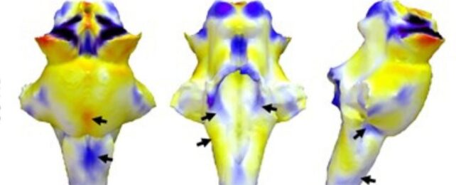
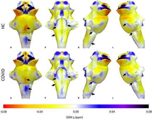
Their brains were scanned at least three months after leaving hospital, yet even then, researchers noticed signs of inflammation in all three parts of the brainstem compared to healthy participants of the same age.
The human brainstem consists of the medulla oblongata, the pons, and the midbrain, all part of the brain's 'automatic control center', which regulates sleep cycles, as well as breathing and heart rates. Fatigue, breathlessness, and elevated heart rates are some of the most common symptoms of long COVID.
"The fact that we see abnormalities in the parts of the brain associated with breathing strongly suggests that long-lasting symptoms are an effect of inflammation in the brainstem following COVID-19 infection," says neuroscientist Catarina Rua from Cambridge.
A similar pattern is found in those who have suffered severe traumatic brain injuries – a patient group with many of the same symptoms.
What's more, autopsy studies on those who have died with long COVID have shown inflamed brainstems with tissue degeneration. This has led some scientists to suspect that the virus is sneaking into the brainstem via the vagus nerve.
In living brains of those with long COVID, however, conventional MRI studies have shown no structural abnormalities in the brainstem.
"Things happening in and around the brainstem are vital for quality of life, but it had been impossible to scan the inflammation of the brainstem nuclei in living people, because of their tiny size and difficult position," explains Rua.
"Normal hospital-type MRI scanners can't see inside the brain with the kind of chemical and physical detail we need. But with 7T (7 Tesla) scanners, we can now measure these details."
Last year, when researchers in Australia used 7T scanners on 8 long COVID patients, they found brainstem regions were significantly larger than those of 10 healthy control subjects. This indicates ongoing brain inflammation.
Notably, that same pattern appears in patients with chronic fatigue syndrome or myalgic encephalomyelitis (CFS/ME) – a condition with similar symptoms that may be related to long COVID or triggered by similar factors.
Earlier this year, researchers in the US also tracked immune cells in a small group of long COVID patients using a PET (positron emission tomography) imaging test. They, too, found abnormal immune activity in the brainstems of patients.
"The brainstem is the critical junction box between our conscious selves and what is happening in our bodies," explains neuroscientist James Rowe from Cambridge.
"The ability to see and understand how the brainstem changes in response to COVID-19 will help explain and treat the long-term effects more effectively."
The study was published in Brain.